A lot depends on PDA size and its effect, especially for the extreme preemie baby and the premature baby with PDA. When trying to understand the effects of PDA in a small baby, there are several factors one looks at:
Clinical Factors
Ventilatory Setting Changes
Systemic Problems: GI, Neuro, Renal
Echo Parameters: those apart from PDA Size include several factors like LA: Ao ratio etc.
Here we are going to discuss the nuances of measuring a PDA. There is no article I could find explaining the details of the same.
VIDEO
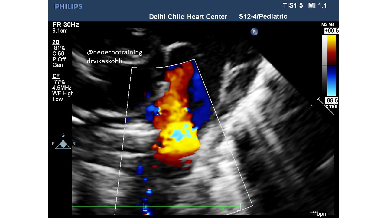
Here if we focus on the PDA flow from Desc Aorta to PA, we will notice the color width appears very variable. That’s the way it looks when we see it at the babi’s heart rate.
So let’s analyze and understand this picture bit by bit.
Fig 1
The jet in this frame looks wide. Let’s just understand the basics here.
Which structures are which?
Let’s have a look in further detail.
Figure 2
The Desc Ao, the MPA (main Pulmonary Artery) and the Right Pulmonary Artery are seen here.
The LPA (left pulmonary artery) is not seen in this view when PDA is seen. This does not happen in all PDA’s and short-axis views. In some, you can see the LPA and the PDA along with the RPA.
So once we have these structures outlined, we should now decide where we want to measure.
Figure 3
3 planes transactionally are marked. A, B, C. A is in the MPA, B is close to where the
PDA flow enters the PA. And C is the exact point between the Descending Ao and the PA. So it is in this plane that we are supposed to measure the PDA. Now let’s adjust the color gain as explained below.
Figure 4
This figure shows color flow across PDA with slightly decreased color gain. Note the diameter of PDA appears lesser than the above pics.
Figure 5
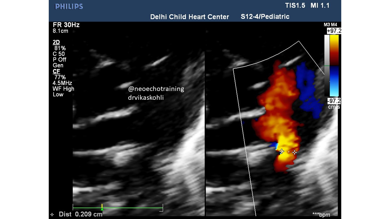
This image shows the same one as above but along with its simultaneous 2D diameter measurement. The diameter measured on color frame is as noted: 2.09 mm.
Figure 6
But on 2D the diameter measures 1.75 mm. This is the exact diameter we need.
In conclusion: the diameter measurement should be followed as follows:
Try to get the systolic frame
Decrease the color gain (not the scale)
Increase the color scale to max
Measure it in color AND
In the same frame press button {HIDE} on Sonosite or {COLOR SUPRESS} on other brand machines and measure the PDA in 2D.
There should be only a small difference between the 2 measurements
The 2D measurement is the true diameter of the PDA
HOW IMPORTANT IS THE DIAMETER
(This is the topic of the Next Newsletter)
Of the 6 studies reviewed for scoring systems of PDA being hemodynamically significant, the diameter of PDA was included in every scoring system. So this is why it is so important.
Next Newsletter…..Scoring Systems of hsPDA


